18 Nov Experts Cut Radiation Exposure in Children Who Need Repeat Brain Scans
A team of experts has developed a way to minimize dangerous radiation exposure in children with conditions that require repeat CT scans of the brain. The experts say they reduced exposure without sacrificing the diagnostic accuracy of the images or compromising treatment decisions.
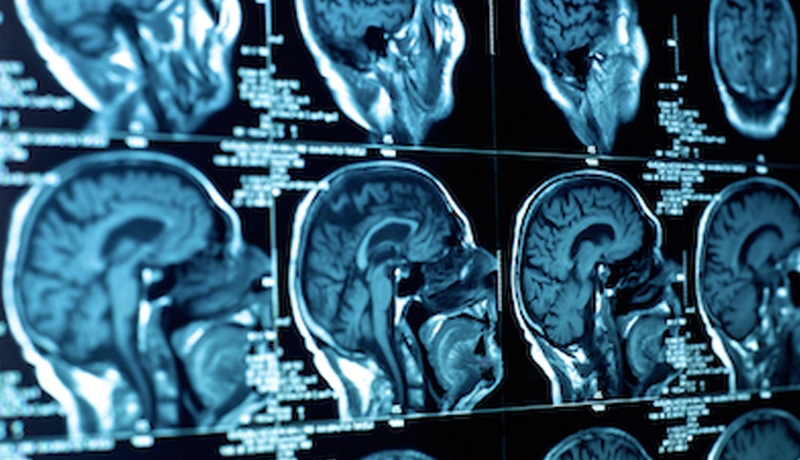
The approach calls for using fewer x-ray snapshots, or “slices” of the brain, taken by CT scanners—from seven instead of 32 to 40 slices. The study, which hit the news in October, 2013 was reported in the Journal of Neurosurgery and on the website Science Daily. The approach used in the study reduced radiation exposure by an average of nearly 92 percent per patient compared with standard head CT scans, while still rendering the images diagnostically accurate.
The study, conducted by a team of pediatric neurosurgeons and neuroradiologists at the Johns Hopkins Children’s Center on 50 patients age 17 and younger, involved analysis of CT scans of patients with hydrocephalus, excessive fluid in the brain, that requires periodic fluid-draining surgeries and a head CT before each procedure.
Hydrocephalus is often treated with ventriculo-shunting, a catheter that drains excess fluid from the brain into the abdomen. Children with brain shunts require periodic imaging to assess catheter position and function. Shunt failure is unfortunately common, and occurs when a catheter dislodges or infection sets in. It is serious and considered an emergency. However, because shunt failure often causes non-specific symptoms such as headache, vomiting and fever, a definitive diagnosis requires a brain scan.
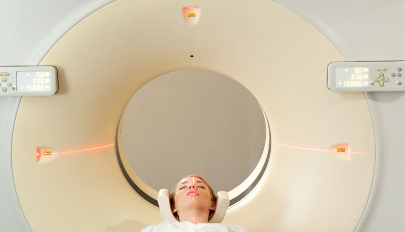
The Hopkins team says the radiation-minimizing technique could be especially valuable in pediatric emergency rooms, where the need for quick diagnosis necessitates CT scans, as opposed to the use of the more cumbersome, radiation-free imaging alternatives like MRI. CT scans can also be used for routine evaluation in smaller community hospitals that may lack MRI equipment, the researchers say.
CT scans are invaluable imaging tools. They have revolutionized the diagnosis and treatment of many conditions by providing fast, reliable and accurate images, but they have also driven up levels of radiation exposure in both children and adults, the study’s investigators say. Ionizing radiation, used in X-rays and CT scans, has been long implicated in the development of certain cancers because of its potential damage to DNA. Children are especially vulnerable to the effects of radiation because of their smaller size, growing tissues and rapidly dividing cells, and because of their longer life-spans which allow slow-growing cancers to present themselves decades after exposure, the researchers note.
Over 62 million CT scans performed each year in the United States, four million of them in children. Experts estimate that radiation from medical imaging is the single largest source of radiation exposure in the population, and that up to two percent of all cancers stem from exposure to medical radiation.
The study’s authors believe that limited-slice CT scans achieve the balance between accurate images and minimizing radiation exposure in children. This is no easy feat, says the senior study investigator.
ANA is a team of expert neurosurgeons and medical professionals, who combine their decades of knowledge to provide information, events, and articles on a range of neurological conditions.