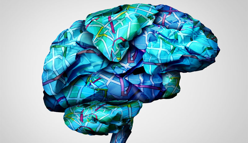
21 Feb What is Brain Mapping and How Is It a Part of Our Future?
In Fall 2010, the National Institutes of Health (NIH) awarded grants totaling $40 million to map the human brain’s connections in high resolution. Better understanding of such connectivity promises improved diagnosis and treatment of brain disorders, an exciting prospect for ANA, and for all of us who work with the brain.
The grants are the first awarded under the Human Connectome Project in support of two collaborative research projects. The first is led by researchers at Washington University, St. Louis and the University of Minnesota, Twin Cities. The other is led by investigators at Massachusetts General Hospital (MGH)/Harvard University, Boston, and the University of California Los Angeles (UCLA).
In Phase 1 (years 1-2, Fall 2010-Spring 2012) data acquisition and analysis methods were optimized for 16 major project components. In Phase II (years 3 – 5, Summer 2012–Summer 2015), data was acquired from a cohort of 1,200 healthy adults, with primary data acquisition at three of the institutions mentioned above. The resulting data is being made publicly available at regular intervals, thereby enabling many explorations and analyses of brain circuitry even as data collection continues.
Altogether, the Human Connectome Project will lead to major advances in our understanding of what makes us uniquely human and will set the stage for future studies of abnormal brain circuits in many neurological and psychiatric disorders.
The Human Connectome Project is an ambitious effort to map the neural pathways that underlie human brain function. The overarching purpose of the Project is to acquire and share data about the structural and functional connectivity of the human brain. In addition, it will also greatly advance the capabilities for imaging and analyzing brain connections, resulting in improved sensitivity, resolution, and utility, thereby accelerating progress in the emerging field of human connectomics. Recent advances in non-invasive neuroimaging have enabled the measurement of connections between distant regions in the living human brain, thus opening up this new field of research: human connectomics—comprehensive maps of the brain’s connections.
Information about brain connectivity is being obtained using two powerful and complementary MR imaging modalities. This is particularly of interest to our community of neurosurgeons, as we rely on the development of new imaging technologies to diagnose conditions and perform brain surgery.
The Human Connectome Project is providing a treasure trove of information at an unprecedented level of detail. Evaluation of this data must respect the profound anatomical and functional complexity of the human brain. The HCP is mapping the human connectome as accurately as possible in a large number of normal adults.
In June, 2014, New York Times reporter Jim Gorman wrote two articles on the stunning experience of participating in the brain mapping study, including an evaluation of his own brain scans. In addition to describing what it is like to be part of the study, both in-scanner and out-scanner, Gorman wrote a more personal philosophical piece on what it is like to be scanned and, ultimately, to be able to look at images of your own brain.
ANA is a team of expert neurosurgeons and medical professionals, who combine their decades of knowledge to provide information, events, and articles on a range of neurological conditions.